Medical Island wins IndiBlogger Awards 2017

Check it Out Here
Abdominal Compartment Syndrome

Abdominal Compartment Syndrome – A Silent but Lurking Danger!

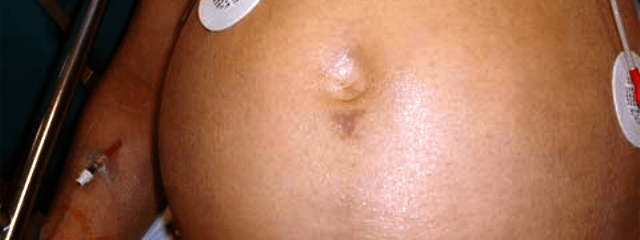
Abdominal Compartment Syndrome and Intra-abdominal Hypertension are two sides of the same coin. While Intra-abdominal Hypertension exists when intra-abdominal pressure exceeds the normal level which is set between 20 to 25 mmHg, Abdominal compartment syndrome is said to exist when intra-abdominal hypertension is accompanied by manifestations of organ dysfunction, which dramatically reduces upon abdominal decompression. The organ systems commonly involved include the pulmonary, cardiovascular, renal, splanchnic, musculoskeletal/integumentary (abdominal wall), and the central nervous system.

Certain population groups at particular risk for Abdominal Compartment Syndrome are:

  • Severe blunt and penetrating abdominal trauma
  • Ruptured abdominal aortic aneurysms
  • Retroperitoneal haemorrhage
  • Pneumoperitoneum
  • Neoplasm
  • Pancreatitis
  • Massive ascites
  • Liver transplantation
  • Massive fluid resuscitation
  • Accumulation of blood and clot
  • Bowel oedema
  • Forced closure of a noncompliant abdominal wall
  • Circumferential abdominal burn eschars

Epidemiology

  • Incidence among those with identifiable risk is 14%, while the incidence following primary closure after repair of ruptured abdominal aortic aneurysm was found to be 4%.
  • In the trauma population, those patients undergoing abbreviated or “Damage Control” laparotomy, particularly those  with intra-abdominal packing are at an increased risk. Interestingly, an open abdomen does not necessarily mean total absence of the risk of this syndrome.

Signs and Symptoms

  • Respiratory Failure that is characterized by impaired pulmonary compliance resulting in elevated airway pressures with progressive hypoxia and hypercapnia. High airway pressures may be needed in this scenario to overcome the high extrathoracic pressure exerted through the diaphragm and not to overcome an intrinsic lung problem. The earliest manifestations of this syndrome may be a subtle pulmonary dysfunction or an elevated peak airway pressure. Elevated hemidiaphragms with a loss of lung volume may be the only sign in an otherwise innocuous patient.
  • Haemodynamic Indicators: Elevated heart rate, hypotension, elevated pulmonary artery wedge pressure and central venous pressure, reduced cardiac output, and elevated systemic and pulmonary vascular resistance. A decreased venous return is central to the pathophysiology of abdominal compartment syndrome.
  • Renal function impairment is manifested as oliguria progressing to anuria with resultant azotemia. It is only partly reversible by fluid resuscitation.
  • Raised intracranial pressure is also another clinical manifestation.

How to Clinically Confirm a Diagnosis of Abdominal Compartment Syndrome

Intra-abdominal Hypertension is present when intra-abdominal pressure exceeds 20 mmHg

  • Direct measurement of intra-abdominal pressure by means of an intraperitoneal catheter
  • Bedside measurement can be achieved by transduction of pressures from indwelling femoral vein, rectal, gastric, and urinary bladder catheters. Of these, measurement of urinary bladder pressure and gastric pressures are the most common and easily accessible.
  • Intragastric pressure measurements are taken from an indwelling nasogastric tube. This varies within 2.5 cm H2O or 1.84 mmHg (conversion factor 1 cm H2O = 0.736 mmHg) of urinary bladder pressures.

In 1984, Kron et al. described  a method to measure intra-abdominal pressure at the bedside with the use of an indwelling Foley Catheter as follows:

  • Sterile water (50 to 100 ml) is injected into the empty bladder through the indwelling Foley catheter.
  • The sterile tubing of the urinary drainage bag is clamped just distal to the culture aspiration port.
  • The end of the drainage bag tubing is connected to the Foley catheter.
  • The clamp is released just enough to allow the tubing proximal to the clamp to drain fluid from the bladder, then reapplied.
  • A 16-gauge needle is then used to Y-connect a manometer or pressure transducer through the culture aspiration port of the tubing of the drainage bag.
  • Finally, the top of the symphysis pubic bone is used as the zero point with the patient supine.

A Viable Conclusion

Abdominal Compartment Syndrome is best prevented and any existing predisposing factor is looked into and prevented if possible. It is an important component in the critically ill patients and those with blunt abdominal trauma. One must remember that renal failure due to abdominal compartment syndrome does not respond to fluid resuscitation and should trigger us to consider it in the differential diagnosis. It is imperative that the clinician remains alert to this silent but lurking danger that is Abdominal Compartment Syndrome.
Exodus of Indian Doctors

Will the Exodus of Indian Doctors Continue?

Despite numerous policies in place to achieve self-sufficiency in the health sector, the number of Indian doctors serving the disparate rural population is drastically declining. Many efforts to drive the tech-savvy, white-collared and highly ambitious doctors to the unkempt peripheries of the country has been an utter failure. India still suffers an acute shortage of about 6,00,000 doctors, one million nurses, 200,000 dental surgeons and large numbers of paramedical staff. So, this issue is not just about Indian Doctors. Is it? Well, no, but I focus here mainly on the doctors’ dilemma.

Are the Doctors to be blamed?

Sadly, the doctors are only to be pitied. I say this because of the indifferent attitude of healthcare policy makers who ignore the changing needs of the evolving doctor community. It is no more the age of the general practitioner, and a simple medical degree is merely looked down as “dismal”. It is a general trend that specialists are sought after, and an MS/MD is the bare minimum for a doctor to practice satisfactorily. It is also true that despite the numerous medical colleges being set up, only a few are sanctioned or equipped to run post-graduate degrees. Hence, there is a huge disparity between the number of medical graduates and the number of post-graduate seats being offered in the country. This signals the clogging of an already overburdened health care system. Newly graduated doctors can only practice as residents in teaching institutes or as petty medical officers with a meagre paycheck that drives most of them crazy. The unfathomable amount of investment of time and money during their medical education only drives these hapless newbies to the brink of desperation. Only a few manage to land PG seats without paying hefty donations to institutes that exist for themselves. It is a sad situation, but a real and obviously debilitating experience for the many medical graduates that come out each year from various medical colleges across India.

An Obvious Solution

India is the biggest exporter of doctors in the world. With an overseas workforce equal to almost 10 percent of the Physicians in India, it is quite a contributor. The reason is obvious – a huge backlog of medical graduates waiting to get into a post-graduate course! With the increasing public awareness about healthcare issues and the demand for specialists and super-specialists, there is an increasing need for more post-graduate institutes that can cater to the backlog, but changes in India take time. So, another rather easy channel is to move out of the country to acquire specialist training from abroad, while others switch over to other allied healthcare areas like administration. This has in one way eased the burden on the post-graduate institutes in India but caused a dearth of doctors in the rural setup.

Many medical graduates from India seek to pursue education abroad or to set up practice abroad. Favoured locations have been UK, US, Canada, Australia, New Zealand and many other places. They provide excellent post-graduate trainings which is a bonus much beyond the financial implications. The recent decision by MCI to recognize foreign degrees attained abroad has indeed encouraged many who have settled abroad to consider coming back to India.

Some interesting stats about doctors of Indian origin:

One out of every 10 practicing physicians in Canada have Indian origin.

40,000 Indian doctors in the UK treat approximately half of its population.

50,000 physicians and approximately 15,000 residents and students in the US are Indians.

20% of doctors in Australia have Indian roots.

Isn’t that significant? Yes, it does. This makes India a formidable medical giant in terms of the sheer number of medical graduates it exports to other countries year after year.

Is there still an Exodus?

The number of Indian doctors abroad keeps increasing every single year despite numerous incidences of racial overtones and injustices toward Indian doctors. Reasons behind this exodus still being a comparatively better financial deal and most often a chance to explore possibilities of higher education. There is still a tendency among young or fresh doctors to consider opportunities abroad. Indian authorities need to sincerely consider a drastic change in health policies to improve the living standard, the emoluments, and an improved system to accommodate doctors serving the rural population. This could possibly improve the outlook among the growing number of junior doctors in India and give them a reasonably satisfying career in India itself.

Will the Exodus continue?

We will have to wait and see. If there are changes in the healthcare system in India with adequate facilities for fresh medical graduates, I should say there might be a change. With India becoming a major player in the medical tourism industry, the trend might change. [box type=”spacer”]We hope to see many more Indian doctors abroad come back to serve India with more vigour and valour.[/box]Indian Doctors' Woes

Acetaminophen Poisoning

Management of Acetaminophen Toxicity

Acetaminophen or commonly known as Paracetamol, is the most used antipyretic and analgesic in the whole world. It is readily available over-the-counter as a component of common drugs for fever and headache in most parts of the world. The chemical name of paracetamol is N-acetyl-p-aminophenol (APAP). Due to its ubiquitous presence, acetaminophen poisoning is a common and sometimes a debilitating consequence.

Pathophysiology of Acetaminophen Toxicity

The maximum recommended daily dose of acetaminophen is 4 g in adults and 90 mg/kg in children. Acute ingestion of 150 mg/kg or approximately 7-10 g in adults results in toxicity. Acetaminophen is rapidly absorbed from the stomach and small intestine and primarily metabolized by conjugation in the liver to nontoxic, water-soluble compounds that are eliminated in the urine. When there is an overdosage, the conjugation in the liver becomes saturated, and excess APAP is oxidatively metabolized by the cytochrome P enzymes to a reactive metabolite, N-acetyl-p-benzoquinone-imine (NAPOI). This metabolite is rapidly conjugated with glutathione and is excreted by the kidney.

An ensuing cascade of oxidative damage, mitochondrial dysfunction, and the subsequent inflammatory response propagate hepatocellular injury, death, and centrilobular (zone III) liver necrosis. Similar enzymatic reactions occur in extra-hepatic organs, such as the kidney, and can contribute to some degree of extra-hepatic organ dysfunction. Production of APAP’s toxic metabolite, NAPQI, in excess of an adequate store of conjugating glutathione, is associated with hepatocellular damage, necrosis, and hepatic failure

Clinical Features

Four phases mark the course of Acetaminophen toxicity. Hepatic and occasionally renal toxicity is manifest only after 24 to 48 hours postingestion. Patients with significant liver damage subsequently develop  multi-system organ failure including haemorrhage, sepsis, ARDS, and cerebral oedema. Death usually occurs 3-5 days after ingestion.

Phase 1 (0-24 h)

  • Asymptomatic
  • Anorexia
  • Nausea or vomiting
  • Malaise
  • Subclinical rise in serum transaminases levels begins at about 12 hours postingestion

Phase 2 (18-72 h)

  • Right upper quadrant abdominal pain, anorexia, nausea, vomiting
  • Continued rise in serum transaminases levels

Phase 3 (72-96 h)

  • Centrilobular hepatic necrosis with continued abdominal pain
  • Jaundice
  • Coagulopathy
  • Hepatic encephalopathy
  • Nausea and vomiting
  • Renal failure
  • Fatality

Phase 4 (4 d to 3 wk)

  • Complete resolution of symptoms
  • Complete resolution of organ failure

Laboratory Investigations

  • Acetaminophen serum concentration
  • Transaminase levels
  • Measures of hepatic function
  • Serum concentrations of NAPQI-protein adducts
  • Electrolytes and creatinine
  • Human chorionic gonadotropin (HCG) in females of childbearing age: Acetaminophen crosses the placenta, and the fetal liver is able to elaborate NAPQI by 14 weeks of gestation.
  • Urinalysis

Lactate and Arterial blood gas with pH 7.3 is a laboratory component predictive of mortality

Treatment

  1. Decontamination with activated charcoal if within one hour of ingestion: 1 g/kg PO or 10 times the amount of drug ingested
  2. N-acetylcysteine blocks the production of toxic metabolites and hence acts as an antidote. It should preferably be given within 8 hours of ingestion but it could still be beneficial if given within 24 hours of ingestion. It can be given per oral, intermittent intravenous and continuous intravenous routes. The recommended standard dose is 140 mg/kg PO, then 70 mg/kg q4h x 17 doses. Drug is continued until encephalopathy is resolved and INR.IV administration is mandated for fulminant hepatic failure, patient unable to take orally, high serum levels at or beyond 8 hours post-ingestion, or a pregnant patient (higher placental delivery prior to first pass metabolism).
  3. Antiemetics like metoclopramide or ondansetron
  4. Medical toxicologist, hepatologist and transplant surgeon may be consulted depending on the findings.

Admit the patient in an intensive care unit for continuous monitoring and shift the patient to the ward once the toxicity has settled down. Follow-up in the OPD should be strictly pursued.

Remember, any poisoning case should be treated as medicolegal until proven otherwise!

Prognosis

With aggressive supportive care, the mortality rate for acetaminophen hepatotoxicity is less than 2%. Patients who survive should be expected to have a return of normal hepatic function.

Lipogram

Lipogram: Is it the weight of fat in grams?

English is a funny language. Indeed it is! You must be wondering what “Lipogram” stands for? My first thought when I saw the word was “must be something related to fat”. What else with all the media attention on all things relating to fat. Well, I was wrong to say the least.

What is it then?

Interestingly, a lipogram is a literary composition which omits a certain letter of the alphabet. Imagine omitting the letter ‘e’. That would be a linguistic challenge.

Variation on the lipogram include verses which omit one letter but contain every other letter of the alphabet. A glaring example of such a lipogram is this anonymous poem which omits the letter ‘e’ in each line.

Bold Nassan quits his caravan,
A hazy mountain grot to scan;
Climbs craggy rocks to spy his way,
Both tax his sight, but far doth stray.

Interesting, huh! Another interesting read is the nursery rhyme, “Mary had a Little Lamb”, rewritten without the letter ‘s’

Mary had a little lamb
With fleece a pale white hue,
And everywhere that Mary went
The lamb kept her in view;
To academe he went with her,
Illegal, and quite rare;
It made the children laugh and play
To view a lamb in there.

[box type=”spacer”]Next time, remember that lipogram is not the weight of your fatty parts in grams![/box]Lipogram

Methanol Poisoning

Management of Methyl Alcohol Poisoning

Methanol or Methyl Alcohol is a denaturant. It is a component of varnishes, paint removers, windshield wipers, anti-freeze solutions, copy-machine fluid and as a solvent.

Acute Poisoning

Acute Methanol Poisoning is usually a consequence of ingestion of cheap, adulterated, illicit liquor. Methanol is very toxic and requires as little as 30 ml of 40% solution to be fatal. Poisoning is mostly stratified among the people of lower socioeconomic status. On ingestion, Methanol is metabolized in the liver to formaldehyde and formic acid by alcohol dehydrogenase. Although both are toxic, formic acid is what causes the more serious delayed effects. It is also readily distributed in the liver, gastrointestinal tract, eyes and kidneys.

Clinical Features

Methanol is rapidly absorbed from the gastrointestinal tract, peak levels reaching within 1-2 hours. Onset of clinical manifestations is variable.

Early manifestations such as nausea, vomiting, headache, vertigo and gastritis are primarily caused by Methanol itself. High serum levels of Methanol (>40 mg/dl) is associated with obtundation, convulsions and coma.

Late manifestations such as visual disturbances, severe metabolic acidosis, seizures, coma and death are common after 30 hours after ingestion. The latent period is longer when Ethanol is ingested concurrently. Ocular toxicity is associated with diminished vision, flashing spots, dilated pupils, optic disc hyperemia, retinal edema and ultimately blindness.

Severe poisoning leads to myocardial depression, bradycardia and shock.

Laboratory Investigations

Apart from arterial blood gases which is essential, serum osmolality and anion gap assist in diagnosis. Renal and Liver function tests should be done and blood Methanol levels measured, if possible. CT scan of the brain may show bilateral putamen necrosis. A retrospective diagnosis of Methanol poisoning is sometimes based on this finding. Diagnosis is essentially clinical, but calculation of the osmolal and anion gap can help assess severity where serum methanol levels cannot be estimated.

Treatment

1. Unabsorbed Methanol should be removed by gastric lavage.

2. Supportive Measures:

  • Correction of Acidosis
  • Control of seizures
  • Maintenance of nutrition
  • Alkalization of urine enhances excretion of formic acid

3. Specific Measures:

  • Administration of Ethanol to saturate alcohol dehydrogenase in the liver, preventing the formation of toxic metabolite, formaldehyde. A 5% solution of Ethanol is prepared and 15 ml/kg is given as loading dose and then 2-3 ml/kg/hour as maintenance dose orally. It can also be given intravenously.
  • Hemodialysis enhances elimination of Methanol and Formic acid and is indicated when Methanol levels exceed 50 mg/dl. In the absence of serum Methanol level measurements, the osmolar gap is useful to assess the indication for and duration of hemodialysis in Methanol-poisoned patients.
  • Folic acid has been used to enhance conversion of formate to carbon dioxide and water. The dose is 50 mg IV 4 hourly for 24 hours.
  • 4-methylpyrazole, an inhibitor of alcohol dehydrogenase, has been successfully used as an alternative to Ethanol and Dialysis.
BP Apparatus

Discovery of Indirect Blood Pressure Measurement

Most medical discoveries have a notable story to tell. So it is with the discovery of Blood Pressure Measurement. This as I feel is one of the most important discoveries in medical science. With the rise in incidence of hypertension, cerebrovascular accidents, and heart failure, it is only apt that I brought out this interesting history about the past.

The Guys who made it possible

Riva Rocci invented the mercury manometer – this led to the dissemination of indirect sphygmomanometry for systolic pressure. Riva Rocci was the pupil of Potain, the great Parisian diagnostician who is attributed with introduction of the sphygmomanometer. Once Korotkoff had discovered the sounds known by his name, for diastolic as well, the history of the discovery of Indirect Blood Pressure Measurement was complete.

Korotkoff was a surgeon in the Czar’s army and not an internist as was believed earlier. Pirogoff, his teacher, had taught him always to auscultate over any area before performing an incision. On one occasion, while auscultating over an artery just as he was releasing a tourniquet, he heard thumping sounds. Abandoning his original scientific problem which was to study posttraumatic arteriovenous fistulas in the surgical dog laboratory, he tried to quantitate the amount of pressure required to make these auscultated sounds appear and disappear. The sounds correlated well with systole and diastole, as could be ascertained by direct inspection of the flow of blood from the distally severed artery of the dog.

Like most revolutionary discoveries, this landmark discovery was no alien to alienation. When Korotkoff first reported his findings in humans, some considered he had gone bonkers! So were many other scientists and innovators.

IBS

Irritable Bowel Syndrome – Symptoms, Causes and Treatments

Irritable bowel syndrome is a chronic functional disorder of the intestines which causes abdominal pain, often accompanied by cramping, bloating, diarrhea and constipation. It is also referred to as spastic colon or mucous colitis. Irritable bowel syndrome is more commonly known by its abbreviated term – IBS.

There are three forms of IBS depending on the symptoms — diarrhea-predominant (IBS-D), constipation-predominant (IBS-C) and IBS with alternating stool pattern (IBS-A). Clinical investigations are going on a new IBS subtype, post-infectious IBS (IBS-PI).

Symptoms of IBS

[box]IBS symptoms include: • Abdominal pain • Discomfort associated with changes in bowel habits • Fewer than three bowel movements a week • More than three bowel movements a day • Hard or lumpy stools • Loose or watery stools • Straining during a bowel movement • Urgency (having to rush to have a bowel movement) • Feeling of incomplete bowel movement • Passing mucus (white material) during a bowel movement • Abdominal fullness, bloating, or swelling • Chronic pelvic pain • Mental stress • Fibromyalgia and various mental disorders • Researchers believe that there are neurological and psychological disorders associated with IBS.[/box]

Causes of Irritable Bowel Syndrome

The exact causes of irritable bowel syndrome are unknown, but there are factors that may aggravate the trouble. Dietary factors and stress don’t exactly cause IBS, but they can aggravate it. Various studies and researches have noted an IBS connection to diet. Physicians feel that the correct diet can provide IBS relief.

Common IBS diet advice includes:

• Eat soluble fiber foods and supplements.
• Substitute dairy products for soy or rice products.
• Be careful with fresh fruits and vegetables that are high in insoluble fiber.
• Eat small amounts in regular intervals to lessen the symptoms of IBS.
• Avoid eating red meat, oily or fatty (and fried) products.
• Avoid taking dairy products (especially when lactose intolerance is suspected).
• Avoid solid chocolate, coffee (regular and decaffeinated), alcohol, carbonated beverages and artificial sweeteners.

IBS Treatment

IBS treatment includes dietary advice and medication. Developing good dietary habits may be the best treatment for irritable bowel syndrome.

Medications can provide instant IBS relief. These may include stool softeners and laxatives in constipation-predominant, and antidiarrheal (loperamide) in diarrhea-predominant IBS. The low dosage of tricyclic and SSRI antidepressants has shown to be the most widely prescribed medications for helping to relieve symptoms of visceral sensitivity (pain) and diarrhea or constipation respectively. Other medications include:

• Alosetron: Alosetron is a selective 5-HT3 antagonist for IBS-D. It is only available for women in the United States under a restricted access program, due to severe risks of side effects if taken mistakenly by IBS-A or IBS-C sufferers.

• Cilansetron: Cilansetron is also a selective 5-HT3 antagonist that is undergoing further clinical studies in Europe for IBS-D sufferers.

• Tegaserod: Tegaserod is a selective 5-HT4 antagonist for IBS-C. It has shown to have an excellent safety profile for relieving IBS constipation and chronic idiopathic constipation.